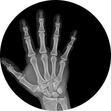
What is arthrogram?
An arthrogram is an X-ray exam of a joint, using a contrast agent and fluoroscopy (a live motion X-Ray). The arthrogram may include additional CT (for cross-sectional views) or MRI (for magnetic resonance) imaging. Though the contrast material may vary, in all cases an arthrogram outlines the structures in the joint and reveals information to the radiologist.
When is an arthrogram used?
An arthrogram is used to reveal the function of cartilage in a joint. Often this procedure is used to image the knee and hip joints, and it is also used when investigating activity in the shoulders, elbows, ankles and wrists. If you have unexplained joint discomfort or pain, or if you have a known issue, such as a tear in the rotator cuff, your doctor may order an arthrogram to diagnose or monitor the problem.
What happens during an arthrogram procedure?
After you are comfortably lying down on the table, the joint to be examined is anesthetized (locally). If fluids are present in the joint, the physician may suction them out. The doctor then injects contrast into the joint, which fills and illuminates the joint in the resulting image. You may be asked to move the joint to circulate the contrast.
The doctor uses fluoroscopy, a kind of live motion X-ray, to monitor the injection and activity of the contrast. If you are having a CT or MRI arthrogram, you will then proceed to that scanner to continue painless imaging of the joint. Afterward, the affected joint should be rested for 12 hours.
What are the risks and benefits of an arthrogram?
An arthrogram is beneficial in that it allows clinicians to get much more clarity on joint health than is provided by a standard X-Ray, which does not show cartilage accurately. MRI, with contrast, accurately shows tears or lesions in cartilage, as well as problems with ligaments, tendons or other joint capsules. It is minimally invasive and presents little risk to the patient.


